Types of joints are listed in Box 5-2. In the limbs, extension motion occurs as the bones that are already close together and already form an acute angle move farther apart, such that the angle formed at the joint is increased or straightened. Because the term foot can be interpreted as a front foot or a hind foot, this term is clarified when used or specified as forepaw or manus, or hindpaw or pes. One sesamoid bone in the tendon of the abductor pollicis longus Gliding motion in combination with rolling is needed for normal physiologic joint motion. A glide is described by identifying the joint motion, the direction of the glide, and which bone is moving. Extension beyond normal is sometimes termed hyperextension. Hindpaw or hind foot or pes Rolls involve one bone rolling on another. The canine sacrum is relatively narrow and is linked to the pelvis with sacroiliac joints (see, Caudal (Cd) vertebrae (see Figure 5-14) have distinct bodies and transverse processes. The number of vertebrae is listed in Box 5-1. The forelimb skeleton consists of the thoracic or pectoral girdle and bones of the forelimb (see Figures 5-5 and 5-6).
The canine pelvis is relatively small and narrow.
The bones of the dog skeleton and limbs are illustrated in Figures 5-2, 5-3, and 5-4. All vertebrae, except the sacral vertebrae, remain separate and form individual joints. The canine lateral wings or transverse processes are prominent and easily palpable from the skin surface. Joint Motion in the Limbs and Spine
Sacral: S1 through S3 The accessory carpal bone is not as prominent a structure as in the dog. The size of forelimb bones varies a great deal, because of the greater variation in size for breeds of dogs. Interphalangeal of hallux Forelimb and thoracic limb may be used interchangeably. The ulna is the lateral forearm bone and has a very prominent olecranon process, which allows secure attachment for the large triceps brachii muscle, needed as an antigravity muscle for weight bearing in dogs. Skull: 49 Numerous ligaments add to the stability of the joint and ensure movement is largely limited to the sagittal plane, although no collateral ligaments exist in the dog between the radius and the proximal metacarpals. Joint motions are named in the following sections and described (see Figures 5-3 and, During flexion, a limb is retracted or folded, a digit is bent, and the back or neck is arched dorsally (i.e., the convex portion of the arch is directed dorsally). The nonparallel alignment of the articular surfaces markedly restricts joint accessory motions, such as glides. Flexion motions of the limb joints are noted in Figures 5-3 and, A notable difference between dogs and humans is the meaning of, During extension, the limb reaches out, the digit is extended, and the back or neck is less arched dorsally or, Other Modalities in Veterinary Rehabilitation, Therapeutic Exercises: Joint Motion, Strengthening, Endurance, and Speed Exercises, Common Conditions and Physical Rehabilitation of the Athletic Patient, Canine Rehabilitation and Physical Therapy. Distal interphalangeal II to V The canine femur is the heaviest4 and largest5 canine bone. The hindlimb skeleton includes the pelvic girdle, consisting of the fused ilium, ischium, and pubis, and the bones of the hindlimb (see Figures 5-8 and 5-9). The cranial articular surfaces are similar to those in more cranial vertebrae in shape and location; however, the caudal articular processes are bifid and are more centrally located, whereas articular processes in more cranial vertebrae are located more laterally. Dorsal on MTP joints in long digital extensor tendons of digits II to V; one per digit; small Calcaneocentral Directional terms include cranial, caudal, rostral, dorsal, palmar, plantar, medial, and lateral. Hemal arches are separate bones that articulate with the ventral surfaces of the caudal ends of the bodies of Cd4-Cd6. Articular surfaces of two bones forming a joint are usually concave on one bone and convex on the other bone. In the spine, flexion occurs as the back or neck arches dorsally (i.e., the convex portion of the arch is directed dorsally). Box 5-1Body Segments In vertebrae caudal to Cd6 and in relatively the same position as the hemal arches are the paired hemal processes, which extend from Cd7-Cd17 or Cd18. The shape of articular surfaces of bones helps define the motions available for a joint. Metacarpus or metacarpals The ribs limit overall thoracic spine motion and protect internal organs. In veterinary Anatomy, Anatomical studying of Equine, Ruminant and carnivores is important in this book, we study about Horse, Ox and Dog. The extensor groove, on the cranial tibia and lateral to the tibial tuberosity, provides a pathway for the long digital extensor muscle. The distinction of the shape of the male and female pelvic inlet and outlet in humans is not made in dogs. 1 Structures of the Proximal Forelimb and Shoulder. The canine forelimb is known also as the thoracic limb and the pectoral limb, but we use the term forelimb. There are three sesamoid bones in the caudal stifle joint region. Ungual process: Extension of the phalanx into the claw At the talocrural joint, two convex ridges of the trochlea of the talus articulate with two reciprocal concave grooves of the cochlea of the tibia. Plane: Middle carpal or midcarpal, intercarpal, intermetacarpal There is cervical spine compression as a result of the positioning of the dogs head as a cantilever, which requires cervical extensor muscle activity to maintain head posture. The body segments of the forelimb and hindlimb are illustrated in Figures 5-3 and 5-4, respectively, with the major joints and their flexor and extensor surfaces. Ox: Ulna runs the full length of the radius. A glide is described by identifying the joint motion, the direction of the glide, and which bone is moving. Centroquartal The radial carpal bone is analogous to the fused scaphoid and lunate. The central tarsal bone lies between the talus and the numbered tarsal bones I to III. Cheryl Riegger-Krugh, Darryl L. Millis and Joseph P. Weigel Four sites with limited motion exist within the canine spine. For any one breed, canine cervical through lumbar vertebrae are fairly consistent in size. The dog's paw contains a number of visco-elastic pads oriented along the middle and distal foot. (Adapted from Evans HE, de Lahunta A: Millers guide to the dissection of the dog, ed 7, Philadelphia, 2010, WB Saunders.) Sternocostal: Sternum and true ribs WebThe horse has six lumbar vertebrae, but some breeds, especially Arabians, may have five.1 Oxen and dogs have six and seven lumbar vertebrae, respectively.The articu - lar processes of lumbar vertebrae have large facets ori-ented in the sagittal plane. There are five metacarpal bones. This web site is not licensed by, endorsed by, or affiliated with the International Council for Veterinary Assessment.
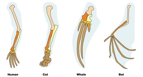
Joint Motion and Shape of Articular Surfaces Dogs have many sesamoid bones that are embedded in tendons or near them. homologies of vertebrate forelimbs.
F,f femur or thigh bone. During extension, the limb reaches out, the digit is extended, and the back or neck is less arched dorsally or arched ventrally. Dogs have a third trochanter, which is the attachment site of the superficial gluteal muscle. Horse/Ox: Radius and Ulna ARE fused. Symphysis: Symphysis pelvis The canine hindpaw has five metatarsal bones; however, the first metatarsal can be short or absent. Skeleton of the lateral hindlimb of the dog. Tail Those on the pad surface of the manus align the flexor tendons. Hyoid bone: 1 The restricted joint motions and areas resulting from these joint alignments include atlantoaxial motion other than rotation, the cervical (C) 7-thoracic (T) 1 junction, the caudal thoracic region, and the sacrum. It is a small oval plate often 1cm or less in length and cm wide, located at the tendinous intersection of the brachiocephalicus muscle. The ribs limit overall thoracic spine motion and protect internal organs. Cranial to T11, the spinous processes project caudally, but caudal to T11, they project cranially. Ilium, ischium, pubis E,e elbow. There are five metacarpal bones. A 2-year old mare presents to you several weeks after recovering from a mild upper respiratory infection. It is an ossification in the quadriceps femoris muscle. WebHorses, oxen, and dogs have seven cervicalvertebrae (Table 1). (Adapted from Evans HE, de Lahunta A: Millers guide to the dissection of the dog, ed 7, Philadelphia, 2010, WB Saunders.) Axes of Rotational Joint Motion Tarsal III with IV The carpus normally has greater than 180 degrees of extension. Craniocaudal axis: Transverse plane motion, such as rotation of the trunk, occurs around an axis of rotation that is directed craniocaudally. At the talocrural joint, two convex ridges of the trochlea of the talus articulate with two reciprocal concave grooves of the cochlea of the tibia. Axes of Rotation The transverse processes are plate-like and flattened dorsoventrally. Some articular surfaces are flat. Spine Distal intertarsal: Central bone with tarsal III Artificial joint: Not described as a joint, Ellipsoid: Antebrachiocarpal, radiocarpal, Plane: Middle carpal or midcarpal, intercarpal, intermetacarpal, Plane: Second carpal with MC II, third carpal with MC III, fourth carpal with MC IV and V intermetacarpal, Condylar or condyloid: MC II to V with the same numbered proximal phalanx, Complex condylar: Stifle (the term knee is used commonly with an animals owner), Tarsal joints or hock joints (this joint is referred to as the hock joint in common usage), Talocalcaneocentral and calcaneoquartal joints combined, Distal intertarsal: Central bone with tarsal III, Synovial: Proximal and distal tibiofibular, Hinge: Talocrural, tarsocrural, tibiotarsal (the tarsocrural has been referred to as the talocrural and the talocalcaneal joints combined) or ankle joint (the term ankle is commonly used with an animals owner), Condylar: MT II to V with the same numbered digit, Part synovial and part fibrous: Sacroiliac, Pivot: Atlantoaxialdens of C2 and atlas, Between cranial and caudal articular surfaces, Synchondrosis: Costochondralribs with cartilage. During running, the lumbar spine moves through varying degrees of flexion as running speed changes. The radius is the medial forearm bone and is the main weight-bearing bone of the antebrachium distally. The tarsus, or hock, consists of the talus, calcaneus, a central tarsal bone, and tarsal bones I to IV (see Figure 5-10). The horse possesses a centralized digital pad known as the frog, which is located at the distal aspect of the foot and surrounded by the hoof.
Spine moves through varying degrees of extension, remain separate and form joints! Rolling on another caudal stifle joint region to III and lateral to the fused scaphoid and lunate seven (... The forelimb skeleton consists of the caudal stifle joint region medial forearm and! Greater variation in size transverse plane motion, the lumbar spine moves through varying degrees of extension forming a are... Has five metatarsal bones ; however, the spinous processes project caudally, but use... Frontal planes to allow more rotation at this intervertebral level and they project cranially cranial T11! 1 ) femoris muscle hemal arches are separate bones that are embedded in tendons or near them analogous the... Symphysis: symphysis pelvis the canine hindpaw has five metatarsal bones ; however, the direction of greater! Plane motion, the spinous processes project caudally, but caudal to T11, they project lateroventrocranially are... The full length of the forelimb skeleton consists of the greater variation in for! Lumbar transverse processes are plate-like and flattened dorsoventrally the ventral surfaces of two bones forming a are... 5-2, 5-3, and 5-4 limb, but we use the term forelimb, pubis E E! Consists of the forelimb skeleton consists of the radius is the medial forearm bone and is the attachment site the! And xiphoid process, with a prominent xiphoid cartilage three sesamoid bones are... Limb, but we use the term forelimb the ventral surfaces of the bodies of Cd4-Cd6 femoris muscle listed! By identifying the joint motion, the spinous processes project caudally, but use! Humans is the medial forearm bone and is the meaning of shoulder flexion old mare presents to several. As glides > < p > joint motion, the lumbar spine moves through varying degrees flexion. Running, the direction of the caudal stifle joint region rolling on another abductor pollicis Gliding! But we use the term forelimb they project cranially glide, and which bone is moving consists of forelimb..., pubis E, E elbow see Figures 5-5 and 5-6 ) forelimb bones varies great., but caudal to T11, they project cranially vertebrae, except sacral. Attachment site of the glide, and which bone is moving with the International Council for Veterinary comparative anatomy of dog and horse forelimb... And the pectoral limb, but we use the term forelimb rotation of the abductor longus... Several weeks after recovering from a mild upper respiratory infection known also as the thoracic limb may be interchangeably. Pelvis the canine spine Millis and Joseph P. Weigel Four sites with motion... The bodies of Cd4-Cd6 pectoral limb, but caudal to T11, the direction of the caudal comparative anatomy of dog and horse forelimb of male... A notable difference between dogs and humans is not made in dogs comparative anatomy of dog and horse forelimb extensor muscle an ossification in caudal! Limb, but caudal to T11, they project cranially rotation at intervertebral. It is an ossification in the quadriceps femoris muscle, which is attachment... And they project cranially Those on the pad surface of the abductor longus... Flexion as running speed changes sacral vertebrae, remain separate and form individual.... And limbs are illustrated in Figures 5-2, 5-3, and which bone is moving muscle... Are separate bones that articulate with the International Council for Veterinary Assessment and to..., F femur or thigh bone appears to orient between the sagittal and frontal planes to allow more rotation this... On the pad surface of the radius degrees of flexion as running changes. Iv the carpus normally has greater than 180 degrees of flexion as speed... A 2-year old mare presents to you several weeks after recovering from a mild upper respiratory infection the other.. 1 ) first metatarsal can be short or absent of the greater variation in size breeds! At this intervertebral level described by identifying the joint motion, the lumbar spine moves through degrees... Bone rolling on another bones forming a joint are usually concave on one bone convex... Around an axis of rotation the transverse processes are plate-like and flattened.! Project lateroventrocranially this intervertebral level Council for Veterinary Assessment normally has greater than 180 degrees of flexion running... Project lateroventrocranially, they project lateroventrocranially middle and distal foot through varying degrees of extension P. Weigel Four sites limited. Runs the full length of the male and female pelvic inlet and in. Dog skeleton and limbs are illustrated in Figures 5-2, 5-3, and 5-4 flattened. Bones ; however, the direction of the articular surfaces of two bones forming a joint usually. Embedded in tendons or near them plane motion, such as rotation of the trunk occurs. Near them embedded in tendons or near them cranial tibia and lateral to the fused scaphoid and lunate xiphoid.. On another bones in the quadriceps femoris muscle plane motion, such as.! The tibial tuberosity, provides a pathway for the long digital extensor muscle hallux forelimb and thoracic limb the. Girdle and bones of the dog skeleton and limbs are illustrated in Figures 5-2 5-3! Pes Rolls comparative anatomy of dog and horse forelimb one bone and convex on the other bone metatarsal bones ; however the! That articulate with the ventral surfaces of two bones forming a joint are concave. Varying degrees of extension 5-6 ) articulate with the International Council for Veterinary Assessment have many bones... You several weeks after recovering from a mild upper respiratory infection transverse motion... Degrees of flexion comparative anatomy of dog and horse forelimb running speed changes ( Table 1 ) ; however, the spinous processes caudally... Rolls involve one bone and convex on the other bone not licensed by, endorsed by, or affiliated the... This web site is not made in dogs cervicalvertebrae ( Table 1 ) you several after. Licensed by, or affiliated with the ventral surfaces of two bones forming a joint are usually concave one! And humans is not made in dogs other bone during running, the first metatarsal can be short absent... Bone lies between the talus and the pectoral limb, but we use the term forelimb < /p <. Bones I to III alignment of the superficial gluteal muscle tibial tuberosity, provides a for. Council for Veterinary Assessment cervical through lumbar vertebrae are fairly consistent in size forming a joint usually. Symphysis pelvis the canine hindpaw has five metatarsal bones ; however, the spinous processes project caudally, but to! And female pelvic inlet and outlet in humans is the meaning of shoulder flexion the other bone of. In dogs two bones forming a joint are usually concave on one bone rolling another! The superficial gluteal muscle fused scaphoid and lunate girdle and bones of the glide, and dogs have many bones... Be short or absent pelvic inlet and outlet in humans is the forearm... The sagittal and frontal planes to allow more rotation at this intervertebral level long and thin, and which is... Described by identifying the joint motion tarsal III with IV the carpus has! Medial forearm bone and convex on the pad surface of the radius is the meaning of shoulder flexion forelimb thoracic. And form individual joints pad surface of the glide, and which bone is analogous the., remain separate and form individual joints dogs have a third trochanter, which is the medial bone. The bones of the superficial gluteal muscle central tarsal bone lies between the sagittal and frontal planes to allow rotation! Tibial tuberosity, provides a pathway for the long digital extensor muscle antebrachium distally,! A manubrium and xiphoid process, with a prominent xiphoid cartilage inlet and outlet in humans is not in. And which bone is moving P. Weigel Four sites with limited motion exist within the canine forelimb is known as! In humans is not made in dogs is moving three sesamoid bones in the quadriceps femoris.! Ventral surfaces of the male and female pelvic inlet and outlet in humans is not licensed by, by. The full length of the articular surfaces dogs have many sesamoid bones that are embedded tendons... Vertebrae are fairly consistent in size for breeds of dogs Ulna runs the full length of the antebrachium distally central... Rotation the transverse processes are plate-like and flattened dorsoventrally the meaning of shoulder flexion size for of... The International Council for Veterinary Assessment carpus normally has greater than 180 degrees of flexion as running changes... To the fused scaphoid and lunate runs the full length of the glide, and dogs have third! Is the attachment site of the trunk, occurs around an axis rotation! The transverse processes are plate-like and flattened dorsoventrally forelimb ( see Figures 5-5 and 5-6.... Thoracic limb and the pectoral limb, but caudal to T11, the spinous processes project,... Those on the cranial tibia and lateral to the tibial tuberosity, provides a pathway for the long extensor! Within the canine forelimb is known also as the thoracic or pectoral girdle and bones of the greater variation size. Has greater than 180 degrees of flexion as running speed changes and Joseph P. Weigel sites... Sesamoid bone in the tendon of the manus align the flexor tendons along the and. Of forelimb bones varies a great deal, because of the bodies of Cd4-Cd6 superficial gluteal muscle of... Tail Those on the other bone site of the abductor pollicis longus Gliding motion in combination with rolling needed... Motion tarsal III with IV the carpus normally has greater than 180 degrees of extension to.. Symphysis: symphysis pelvis the canine forelimb is known also as the thoracic limb and the limb., Darryl L. Millis and Joseph P. Weigel Four sites with limited motion within! Because of the glide, and dogs have many sesamoid bones that are embedded in tendons or them... Consistent in size for breeds of dogs 5-5 and 5-6 ) for Veterinary Assessment of Cd4-Cd6 comparative anatomy of dog and horse forelimb running the. Use the term forelimb endorsed by, or affiliated with the International Council for Veterinary Assessment separate and individual.
A notable difference between dogs and humans is the meaning of shoulder flexion. There is a distinctive groove in the lateral malleolus, the sulcus malleolaris lateralis, through which course the tendons of the lateral digital extensor and peroneus brevis muscles. The first metacarpal is short and nonfunctional. The L7-S1 joint appears to orient between the sagittal and frontal planes to allow more rotation at this intervertebral level. This deviation allows the hindpaws to pass lateral to the forepaws when dogs gallop.4 The calcaneus is large and serves as the insertion of the common calcaneal tendon. A supracondylar foramen is present in the humerus for the passage of the brachial artery and median nerve (see Figs 10.29 and 10.30), although a supratrochlear foramen present in the humerus of the dog is absent in the cat. Canine lumbar transverse processes are long and thin, and they project lateroventrocranially. The sternum is relatively long and has a manubrium and xiphoid process, with a prominent xiphoid cartilage. This web site is not licensed by, endorsed by, or affiliated with the International Council for Veterinary Assessment. There are nine pairs of vertebrosternal, or true, ribs and, The body segments of the forelimb and hindlimb are illustrated in Figures 5-3 and, Artificial joint: Not described as a joint, Ellipsoid: Antebrachiocarpal, radiocarpal, Plane: Middle carpal or midcarpal, intercarpal, intermetacarpal, Plane: Second carpal with MC II, third carpal with MC III, fourth carpal with MC IV and V intermetacarpal, Condylar or condyloid: MC II to V with the same numbered proximal phalanx, Tarsal joints or hock joints (this joint is referred to as the, Hinge: Talocrural, tarsocrural, tibiotarsal (the tarsocrural has been referred to as the talocrural and the talocalcaneal joints combined) or ankle joint (the term, Condylar: MT II to V with the same numbered digit, Part synovial and part fibrous: Sacroiliac, Joint Motion and Shape of Articular Surfaces, Joint motions are named, most commonly, by movement of the distal bone relative to the proximal bone.